Researchers Identify Molecule in Gut that Inhibits Campylobacter Growth
Need to Know
- A naturally occurring substance produced by gut bacteria, called indole, was found to inhibit the growth of Campylobacter jejuni
- Intestinal inflammation reduced indole levels in mice, weakening a natural defense that helped keep C. jejuni growth in check
- The findings could support future development of non-antibiotic approaches for controlling Campylobacter, although further research is needed
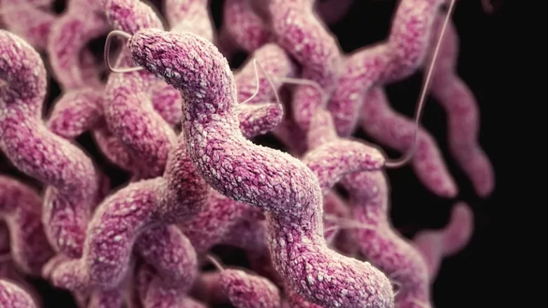
Researchers at Michigan State University (MSU) have identified a gut microbiota-derived metabolite that inhibited the growth and colonization of Campylobacter jejuni, pointing to a potential avenue for developing non-antibiotic interventions against the foodborne pathogen.
The study, published in Science Advances, showed that indole, a molecule produced when certain gut bacteria metabolize the amino acid tryptophan, suppressed C. jejuni by disrupting bacterial respiration and energy metabolism. In mice, administration of indole reduced C. jejuni colonization by approximately 1,000-fold, while an indole-producing probiotic reduced colonization by approximately 100-fold.
“Campylobacter is the most common cause of bacterial foodborne illness in the U.S., but we still know much less about it than we do about other gut pathogens,” said Victor DiRita, Ph.D., Rudolph Hugh Endowed Professor and Chair of MSU’s Department of Microbiology, Genetics, and Immunology, who led the study. “This study gives us a better understanding of the mechanisms that allow Campylobacter to cause disease, but what’s particularly interesting is that we identified a molecule produced by the gut microbiome that can inhibit its growth.”
Inflammation Creates Conditions Favorable to C. jejuni
Building on previous work showing that intestinal inflammation supported C. jejuni growth in ferrets, the researchers developed a mouse model using short-term treatment with dextran sodium sulfate (DSS) to temporarily induce intestinal inflammation. Conventional mice are typically resistant to C. jejuni colonization unless their microbiota or inflammatory pathways are altered.
By three days after infection, DSS-treated mice carried approximately 109–1010 colony-forming units (CFU) of C. jejuni per gram of colon tissue, while the pathogen was undetectable or below the limit of detection in infected mice that did not receive DSS. Infection also exacerbated inflammation and intestinal tissue damage in DSS-treated mice.
Analysis of the gut microbiota showed that DSS treatment depleted several groups of obligate anaerobic bacteria associated with short-chain fatty acid and indole production. Mass spectrometry confirmed significantly lower colonic indole concentrations in DSS-treated mice, while tryptophan accumulated, suggesting reduced microbial conversion of tryptophan to indole.
“We found a significant decrease in the bacteria that produce indole, so we wondered whether indole levels might be lower as well,” said MSU Academic Research Specialist Ritam Sinha, Ph.D., the study’s first author. “When we analyzed indole in the mouse gut, we confirmed this hypothesis.”
Looking for quick answers on food safety topics?
Try Ask FSM, our new smart AI search tool.
Ask FSM →
Indole Disrupts C. jejuni Respiration and Metabolism
The researchers then exposed C. jejuni to indole concentrations of 0.25–1 millimolar (mM), which the study described as physiologically relevant to the gut. Little effect was observed at 0.25 mM, but 0.5 and 1 mM significantly inhibited growth. At 1 mM, C. jejuni showed a near-total loss of viability after 30 hours.
RNA sequencing provided insight into how indole exerted this effect. Exposure to 0.5 mM indole reduced transcripts involved in aerobic and nitrate respiration, lactate utilization, the tricarboxylic acid cycle, and the acetate switch, an important ATP-generating pathway. Respiratory activity decreased approximately tenfold, while intracellular ATP levels fell approximately threefold. Indole exposure also lowered C. jejuni intracellular pH from approximately 7.2 to 6.
Together, the findings suggested that indole interfered with the pathogen's ability to generate and use the energy necessary for growth. The importance of these metabolic pathways was reinforced in mice, where C. jejuni mutants deficient in lactate uptake, nitrate respiration, or the acetate-generating ackA/pta pathway exhibited approximately 100-fold or greater fitness defects relative to wild-type bacteria.
Indole-Producing Probiotic Reduced Campylobacter Colonization
To test whether microbial production of indole could provide similar protection, the researchers used the probiotic Escherichia coli Nissle 1917 (EcN), which produces indole from tryptophan.
Wild-type EcN reduced C. jejuni colonization in mice by approximately 100-fold compared with an EcN mutant unable to produce indole. The mutant provided no comparable protection, supporting the researchers' conclusion that probiotic-derived indole contributed to colonization resistance.
Findings are First Step Toward Non-Antibiotic Treatments
The findings could eventually inform approaches involving indole, indole-producing microorganisms, or other methods of supporting microbiome-mediated resistance to C. jejuni. However, the work remains preclinical, and the researchers noted an important unanswered question: If indole is naturally present in the human intestine, how does C. jejuni overcome this protection during infection?
“We’re still doing a lot of basic research, but that is the foundation for any new therapeutic approaches that we or others might develop,” Dr. DiRita said. “With increasing levels of antibiotic resistance in Campylobacter, this work is pointing us toward innovative, non-antibiotic ways of controlling infection.”